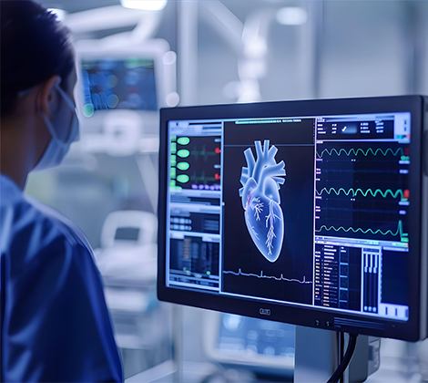
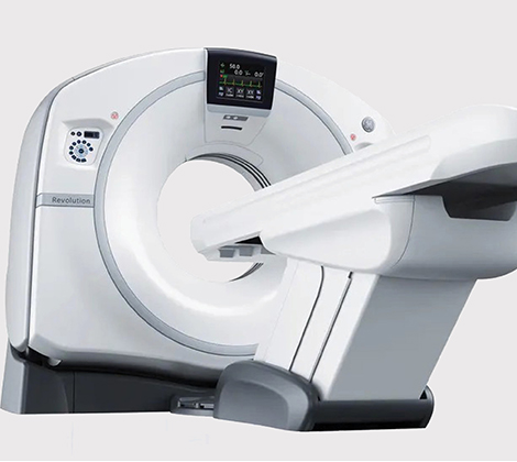
MedCell

Cardiac CT

Our highly specialized exam includes the Computerized Tomography Coronary
Angiogram (CCT). It is applied to look at the arteries that supply blood to the heart
through taking images of the heart and its blood vessels aiming to diagnose the heart
condition.

It detects the narrowed or blocked arteries in the heart (coronary artery disease-CAD) -which remains the most common cause of morbidity and mortality around the world. Moreover, this exam can help the doctor check any other abnormal heart conditions as well.

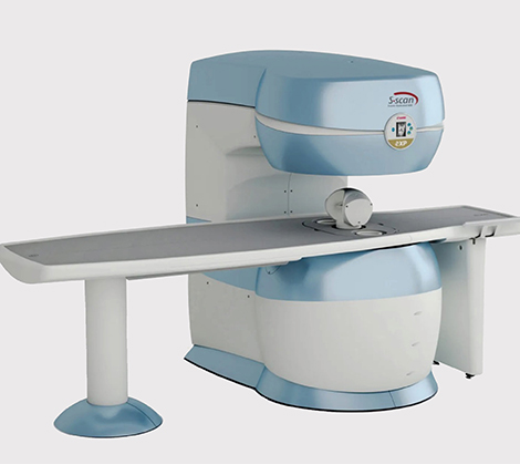
Cardiac MRI for Your Heart Viability


Cardiac MRI is considered to be the most effective examination method that include different studies.

Overall, the tests results will guide the treatment needed for e.g. validate the suitability for procedures such as bypass surgery will be determined based on the findings.

The advantages of Cardiac MRI include:

  • Heart blood pumping measurement
  • Cardiac injuries detection, particularly those caused by heart attacks
  • Identifying the thickness of heart muscle involved
  • Quantitative measurement of blood supply of the cardiac muscle units
  • Identifying if there are any parts of the heart muscle with inadequate blood supply

Prenatal Health – 4D Prenatal Ultrasound

At IMAGES we offer 4D prenatal ultrasounds where amazing, realistic images of the baby is generated by showing movement like a real time video would! Using this technology lets us manipulate the light source and generate images of your little ones.

IMAGES GO

IMAGES offers a mobile X-ray imaging at home service to simplify the medical care of patients whose medical conditions prevent them from visiting our center. When you opt for this service, highly qualified technicians conduct the diagnostic X-ray at your most comfortable setting, your home. This service is fast, safe, and relatively cheaper than other alternatives.

Bone Mineral Density Test

The Bone Mineral Density test assesses your bones’ strength. It is usually used to detect the risk of osteoporosis and bone breakage caused by the decrease of bone density. It is also used to monitor the effectiveness of osteoporosis treatment.

DXA is done for both men and women at risk of osteoporosis.

This test is recommended for people, especially women, at high risk of osteoporosis including those of strong family history, people who have

experienced multiple fractures and falling, smokers, sedentary life-style people, alcoholic, people with hormonal disorders and also people who do not practice sports.

Ultrasound and Doppler

This technology is noninvasive and is used to examine the body’s major organs and tissues intending to evaluate abnormalities, particularly in abdominal, pelvic, sports / muscle injuries as well as Vascular (Doppler) where areas of narrowing or blockage in blood vessel are checked.

It is providing comprehensive and valuable diagnosis of multiple diseases and conditions; thus, anticipating adjacent treatment plans.

Digital X-ray

An X-ray is the most accessible form of imaging. It sends radiation through the body, high levels of calcium parts / parts with high density blocks the radiation, causing them to appear clearly white on the image (Mainly bones). Hence the scanning of latter represents the majority of X-ray examinations.

X-rays are ideal for spotting:

  • Fractures and Dislocations
  • Narrowed Joint Spaces
  • Misalignments
  • Bone Tumors and Bone Cancers

Mammography

A Mammography or mammogram is a safe X-ray scanning technique where the patient is exposed to a small amount of radiation. The goal is to detect cancer (if any) in the breast tissue. The images may show a small tumor that cannot be felt during a regular examination.

This test can be done without any symptoms as its aimed to detect any presence of a cancer at early stage.

If patient comes with specific symptoms, the second test should be applied which is called the “Diagnostic Mammogram – Digital Mammography”, it is more sophisticated compared to regular screening as it converts X-rays into pictures / images of the breasts & Computer Aided Detection looks out for abnormal areas of density, calcification and mass.

CT-Scan

A computed tomography uses a combination of X-rays to create multiple images, known as “slices” from different angles. It offers much higher level of detail compared to regular X-ray. Computerized, 360-degree views of the body structure.

Doctors decide either to use CT or MRI based on medical reasons behind having the scan. Level of details needed for the images is a key factor. However, CT scan is more preferred for specific examinations, those relevant to: Head trauma, internal injuries & bleeding, blood clots, tumors, complex bone fractures & others.

With its 1-3 minutes exam duration, CT scan is an ideal scanning solution for emergency cases.

Open MRI

– Comfortable Non-Claustrophobic O-Scan

O-scan offers an open-machine for maximized patient’s comfort. It features a very compact magnetic field where the patient sits freely on a separate chair. It covers all limbs and joints such as the knee, calf, ankle, foot, hand, wrist, forearm, and elbow.

– S-Scan: The MSK Specialists’ Solution

O-scan offers an open-machine for maximized patient’s comfort. It features a very compact magnetic field where the patient sits freely on a separate chair. It covers all limbs and joints such as the knee, calf, ankle, foot, hand, wrist, forearm, and elbow.

MRI 3 TESLA

IMAGES is proud to install the latest and fastest imaging technology, MRI 3
Tesla. Its powerful magnetic field results in fast, clear, and comprehensive
images.

How is the 3T MRI Better than the Standard One?